Back to Network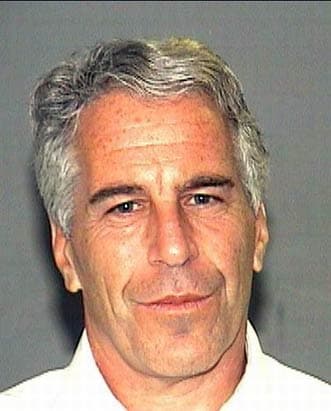

4
Shared Docs
4
Same-Page
4 / 4
Mentions
Document Mentions (4 shared documents)
2 mentions1 same-page
Page ?Same page
HEART x 2 (#2): Myocardial and perivascular fibrosis. Slightly hypertrophied myocytes. LUNG x 5 (#3,4): Slight patchy emphysema. LIVER x 1 (#1): Steatosis involving approximately 50% of hepatic parenchyma. Slight periportal fibrosis. Marked congestion. KIDNEY x 1 (#1): Arteriolar sclerosis. Congestion
eres reveal normal cortical ribbon, white matter, and deep gray nuclei. There is no midline shift. Hippocampi are symmetrical and of normal volume. Ventricles are normal in size and appearance. Axial sections of the brainstem and cerebellum en bloc show the substantia nigra and locus ceruleus to be well-p
2 mentions1 same-page
Page ?Same page
6 HEART x 2 (#2): Myocardial and perivascular fibrosis. Slightly hypertrophied myocytes. LUNG x 5 (#3,4): Slight patchy emphysema. LIVER x 1 (#1): Steatosis involving approximately 50% of hepatic parenchyma. Slight periportal fibrosis. Marked congestion. KIDNEY x 1 (#1): Arteriolar sclerosis. Congestion
eres reveal normal cortical ribbon, white matter, and deep gray nuclei. There is no midline shift. Hippocampi are symmetrical and of normal volume. Ventricles are normal in size and appearance. Axial sections of the brainstem and cerebellum en bloc show the substantia nigra and locus ceruleus to be well-p
2 mentions1 same-page
Page ?Same page
HEART x 2 (#2): Myocardial and perivascular fibrosis. Slightly hypertrophied myocytes. LUNG x 5 (#3,4): Slight patchy emphysema. LIVER x 1 (#1): Steatosis involving approximately 50% of hepatic parenchyma. Slight periportal fibrosis. Marked congestion. KIDNEY x 1 (#1): Arteriolar sclerosis. Congestion
eres reveal normal cortical ribbon, white matter, and deep gray nuclei. There is no midline shift. Hippocampi are symmetrical and of normal volume. Ventricles are normal in size and appearance. Axial sections of the brainstem and cerebellum en bloc show the substantia nigra and locus ceruleus to be well-p
Sponsored
2 mentions1 same-page
Page ?Same page
HEART x 2 (#2): Myocardial and perivascular fibrosis. Slightly hypertrophied myocytes. LUNG x 5 (#3,4): Slight patchy emphysema. LIVER x 1 (#1): Steatosis involving approximately 50% of hepatic parenchyma. Slight periportal fibrosis. Marked congestion. KIDNEY x 1 (#1): Arteriolar sclerosis. Congestion
eres reveal normal cortical ribbon, white matter, and deep gray nuclei. There is no midline shift. Hippocampi are symmetrical and of normal volume. Ventricles are normal in size and appearance. Axial sections of the brainstem and cerebellum en bloc show the substantia nigra and locus ceruleus to be well-p
Sponsored
Mutual Connections (99)
Entities connected to both Steatosis and Ventricles
NEUROPATHOLOGY
ORGANIZATION5 w/ Steatosis·6 w/ Ventricles
Jeffrey Epstein
PERSON5 w/ Steatosis·6 w/ Ventricles
LC-MS
ORGANIZATION4 w/ Steatosis·6 w/ Ventricles
Positive
LOCATION4 w/ Steatosis·6 w/ Ventricles
New York Office of Chief Medical
ORGANIZATION4 w/ Steatosis·6 w/ Ventricles
FUBINACA
ORGANIZATION4 w/ Steatosis·6 w/ Ventricles
HI
Hippocampi
PERSON4 w/ Steatosis·6 w/ Ventricles
AMB-CHMINACA
ORGANIZATION4 w/ Steatosis·6 w/ Ventricles
Special Housing Unit
ORGANIZATION4 w/ Steatosis·6 w/ Ventricles
NA-FUBIC
ORGANIZATION4 w/ Steatosis·6 w/ Ventricles
BS
Barbara A. Sampson
PERSON5 w/ Steatosis·5 w/ Ventricles
Ghislaine Maxwell
PERSON5 w/ Steatosis·5 w/ Ventricles
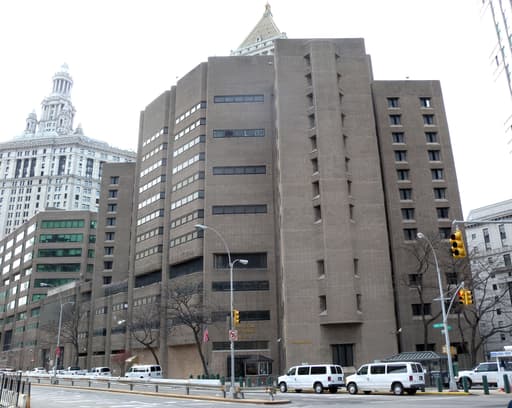
Metropolitan Correctional Center
ORGANIZATION4 w/ Steatosis·5 w/ Ventricles
AMB-FUBINACA
ORGANIZATION4 w/ Steatosis·5 w/ Ventricles
New York Criminal Procedure Law Section 190.30(2
ORGANIZATION4 w/ Steatosis·5 w/ Ventricles
MD-Ph.D.
ORGANIZATION4 w/ Steatosis·5 w/ Ventricles
Horsham
LOCATION3 w/ Steatosis·5 w/ Ventricles
NYPD Phone
ORGANIZATION3 w/ Steatosis·5 w/ Ventricles
PCLAS
ORGANIZATION3 w/ Steatosis·5 w/ Ventricles
HEPATIC
ORGANIZATION4 w/ Steatosis·4 w/ Ventricles